-

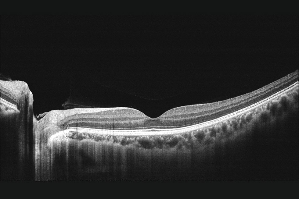
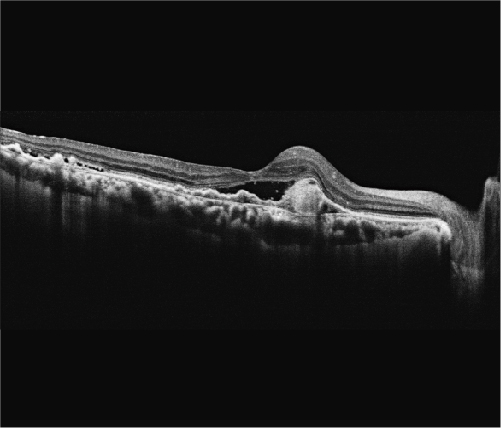
Superior OCT image quality with up to 100 times averaging
Mocean 4000 captures 100 images in less than one second, and merge them together to create a high definition image with minimized speckle noise.
5 μm axial optical resolution (3 μm digital).
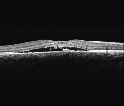
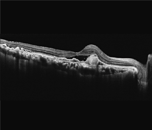
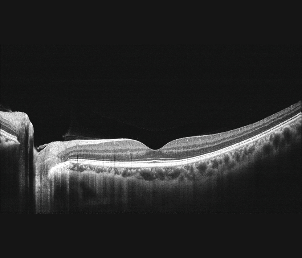
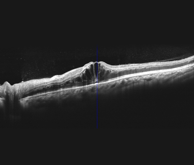
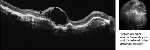
High definition OCT imaging reveals hidden pathological changes.
Diabetic Retinopathy (DR)
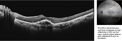
Wet AMD
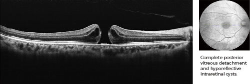
Macular Hole
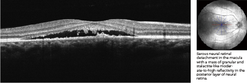
Central Serous Chorioretinopathy (CSC)
-
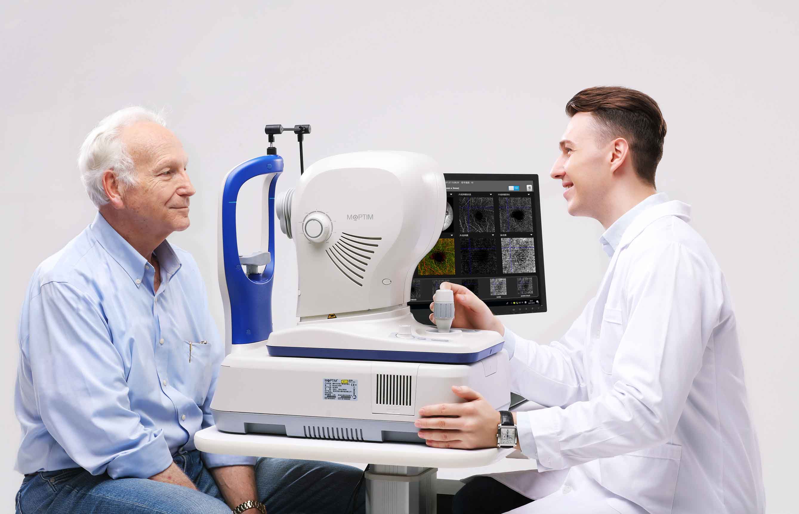
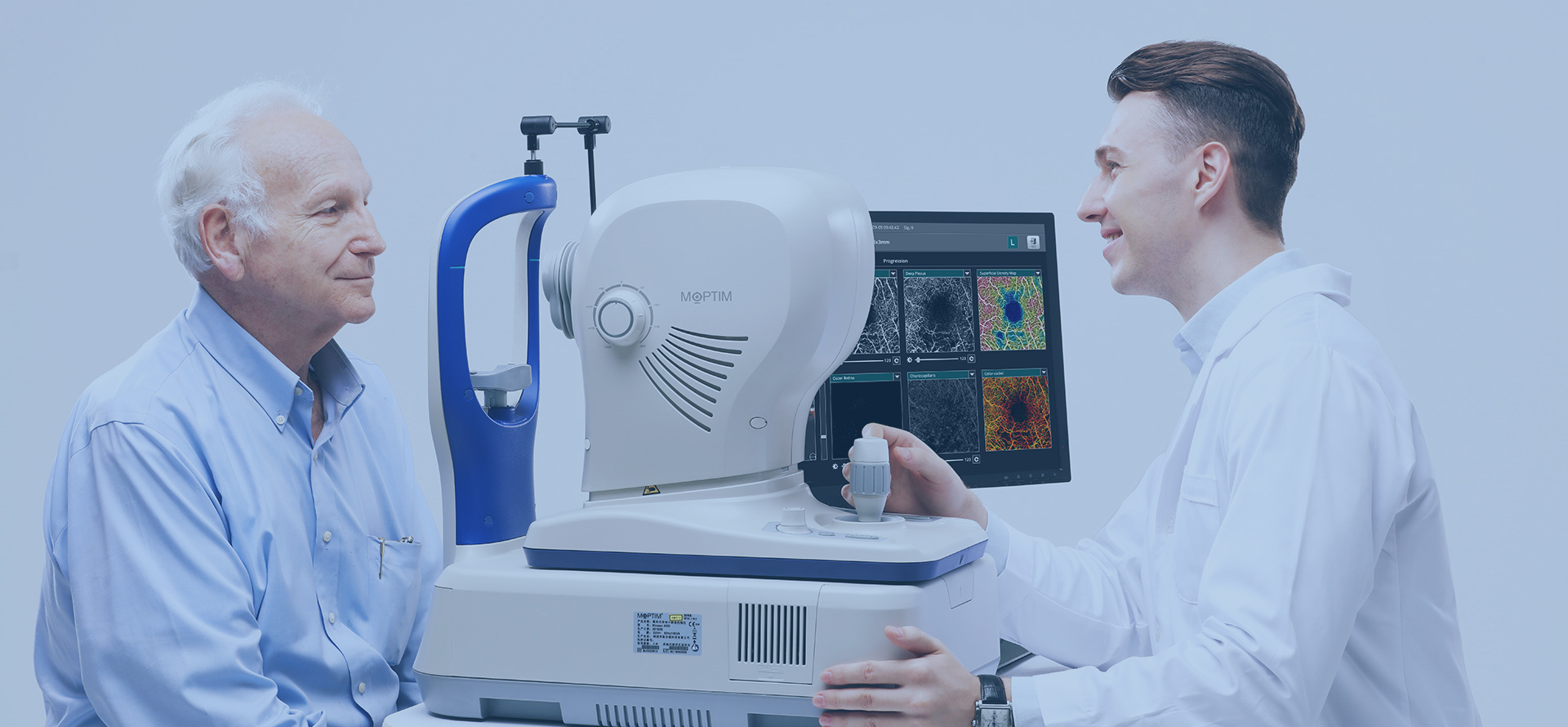
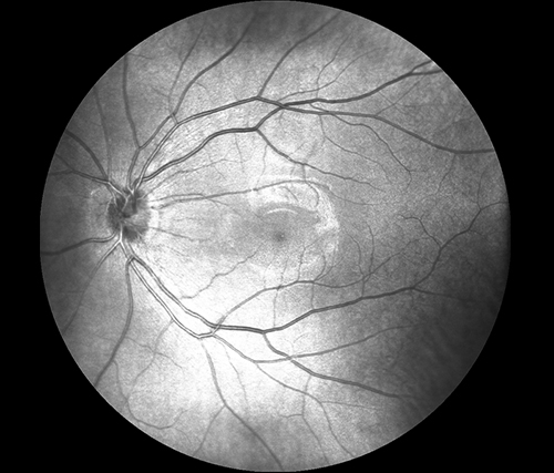
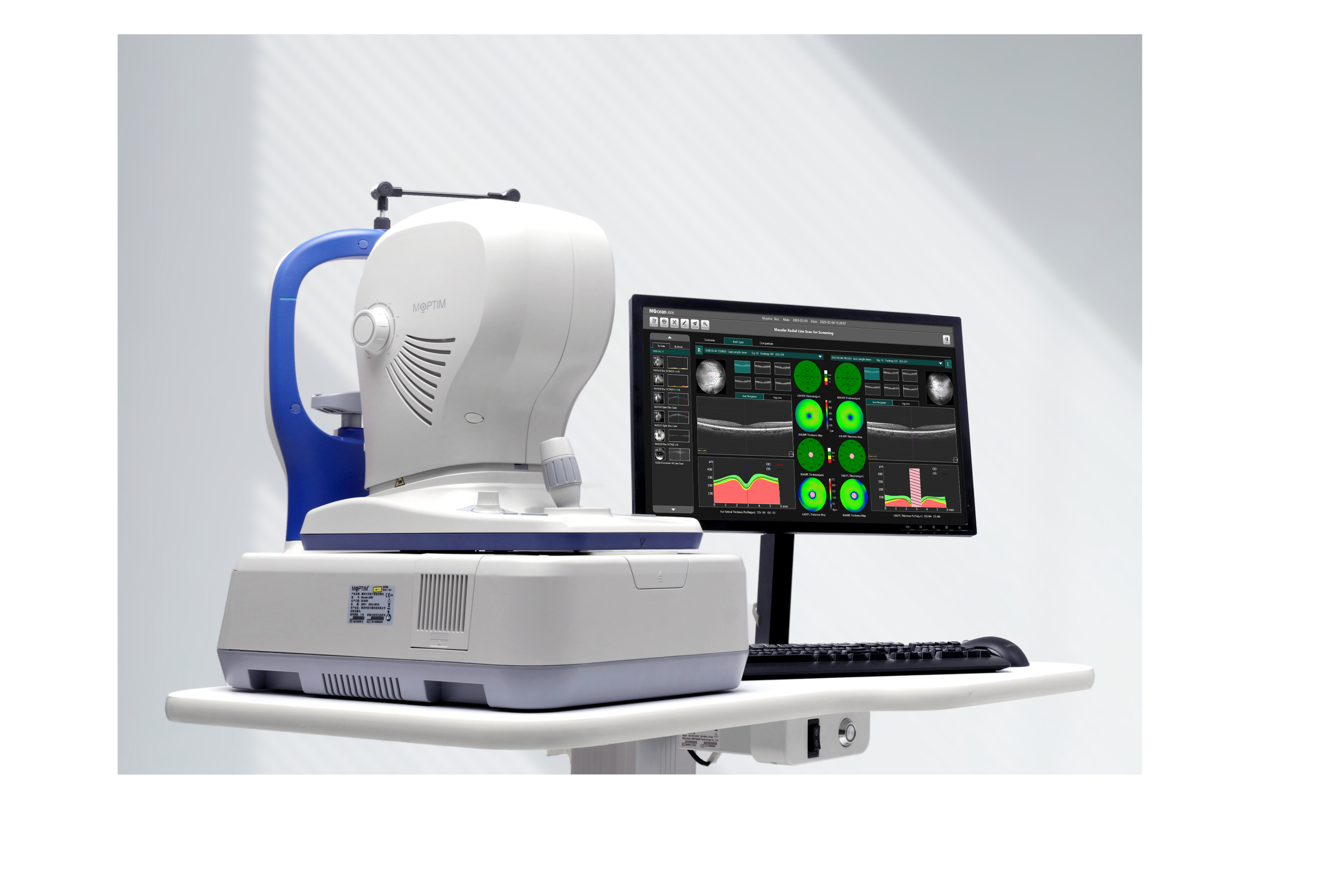
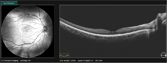
High quality real-time SLO + Eye tracking
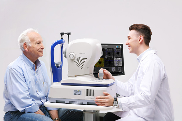
The key advantage of Mocean® 4000 system is the simultaneous acquisition of cross-sectional OCT imaging and 45 degrees fundus imaging based on Scanning Laser Ophthalmoscope (SLO). It gives you an overview of the retina so you can easily locate the lesion area before acquisition. Moreover, the system captures up to 50 SLO fundus images within one second in order to generate an HD fundus imaging.
To minimize the artifacts caused by eye drift and micro saccades, Mocean® 4000 uses SLO-based eye tracker. It performs 100 times tracking per second with 10 microns tracking accuracy and more than 95% success rate, which gives you more confidence in practice.enhanced signal-to-noise ratio.
-
High scanning speed at 80,000 A-scans/s
80,000 A-scans/s allows 1.5s examination for high definition scan, and 1s for 3D scan.
-
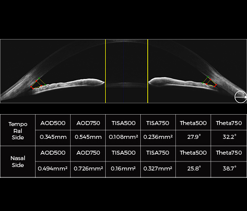
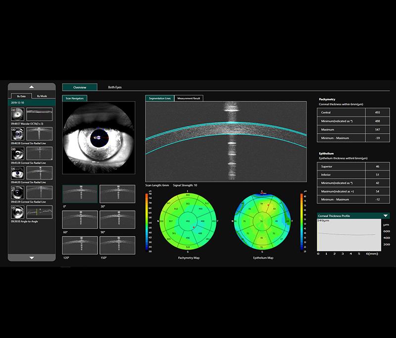
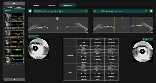
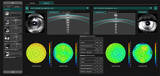
16mm angle-to-angle analysis
16mm angle-to-angle anterior scan with data analysis.
Precise follow-up analysis
-
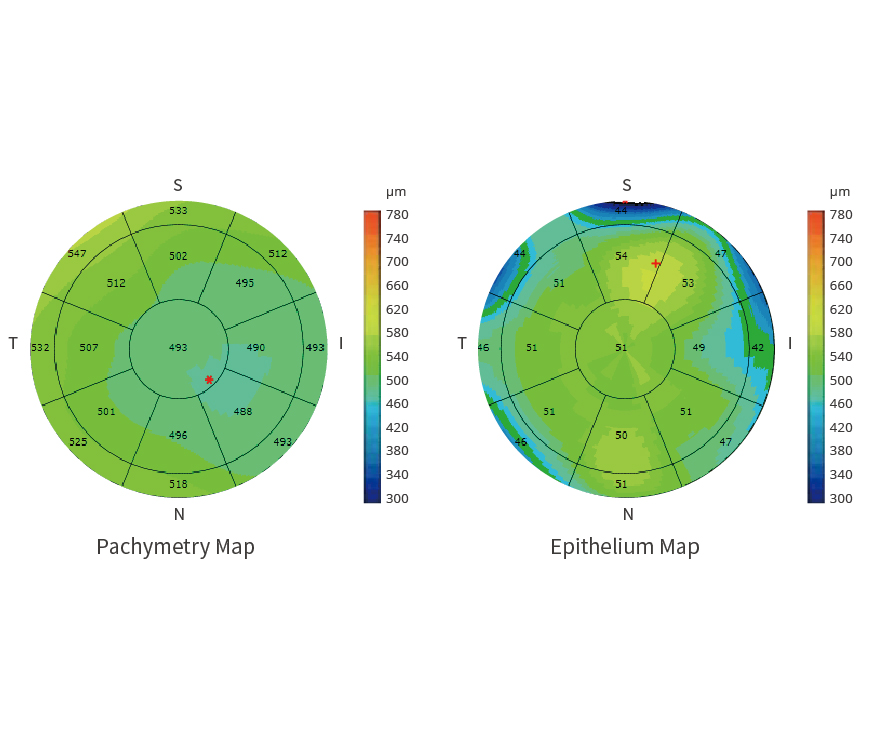
Epithelial thickness mapping
Provides 6mm diameter cornea epithelium thickness map, which is an important part of diagnostics
in refractive surgery, with many important clinical applications.
Both eyes
-
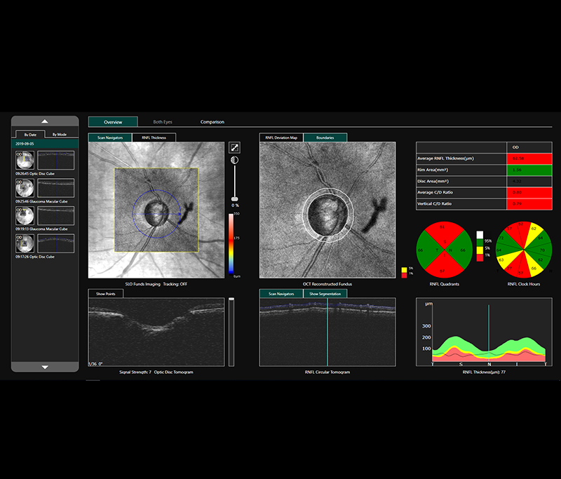
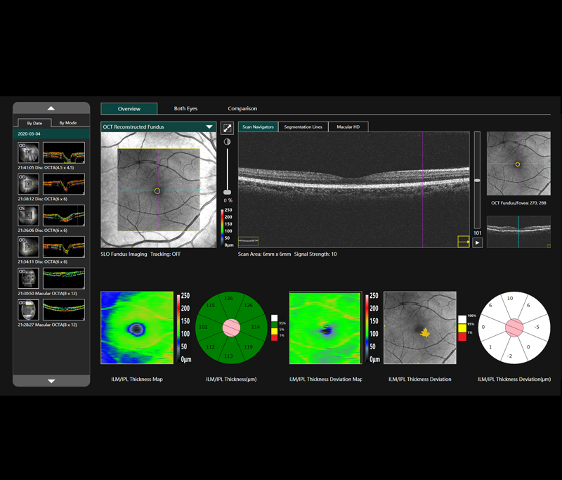
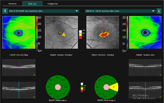
Comprehensive GLAUCOMA analysis (ONH/GCC)
For comprehensive glaucoma analysis, Mocean 4000 offers two scan patterns, glaucoma cube scan in macular area for GCC analysis and glaucoma cube scan in disc area for ONH analysis. Evenly distributed sampling point with 200 x 200 A-scans provides reliable information for early glaucoma detection and management.
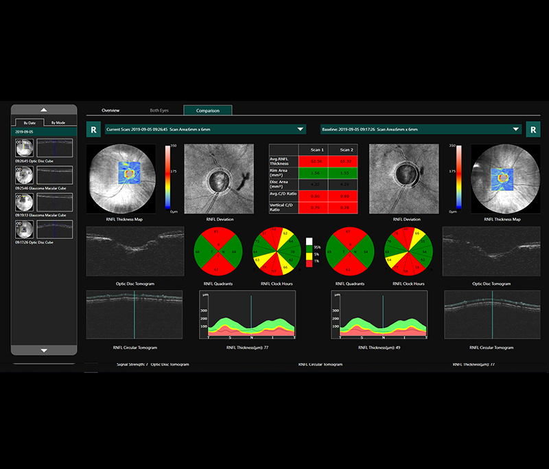
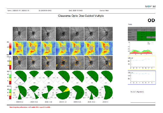
ONH comparison

GCC both eyes
Progression analysis
-
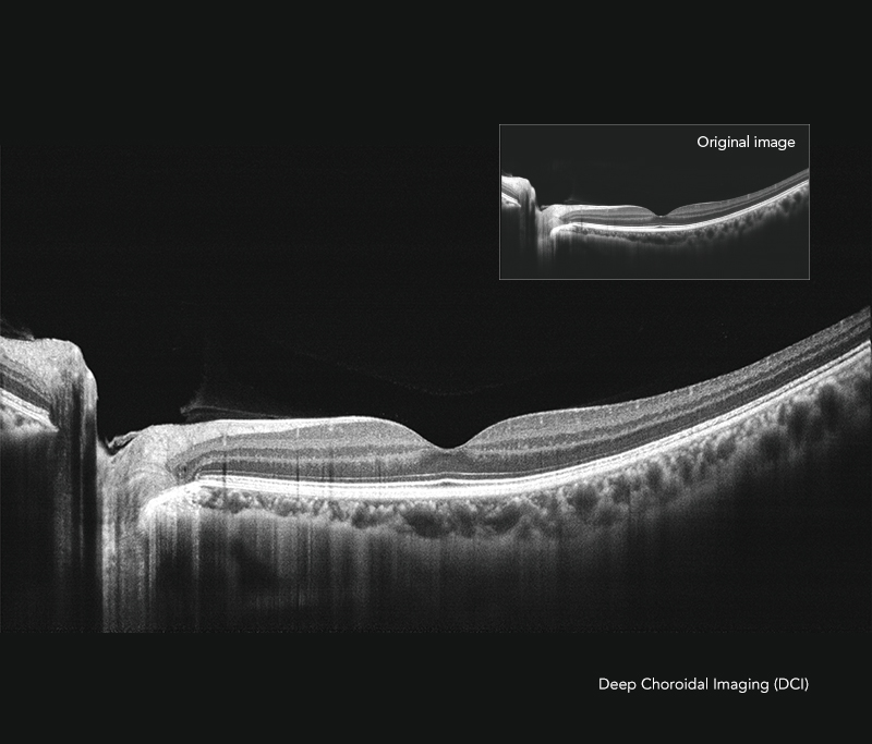
Deep Choroidal Imaging (DCI) mode
Using Deep Choroidal Imaging for detection of choroidal neovascularization.
-
Comprehensive software analysis and free upgrade
The Mocean® 4000 system provides 9 scan patterns to help you improve diagnostic efficiency:
Retina (HD line, Six-Radial lines, Multi, 3D Cube)
Glaucoma (Glaucoma Disc for ONH analysis, Glaucoma Macular for GCC analysis)
Cornea (HD line, Six-Radial lines, Angle-to-Angle)
The software analysis features are always up-to-date and free for upgrade (excluding OCTA module).
-
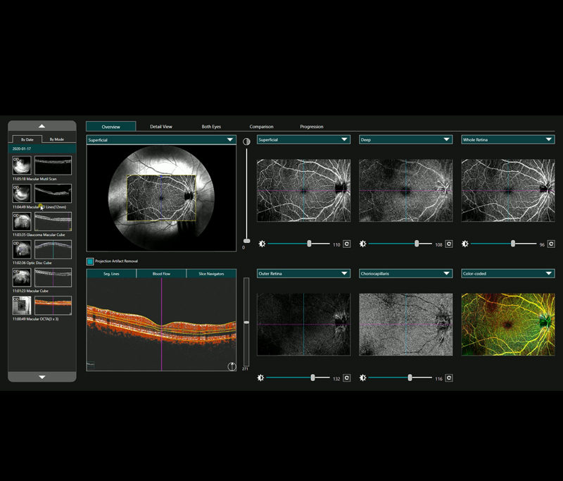
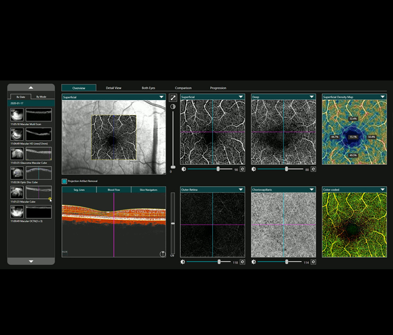
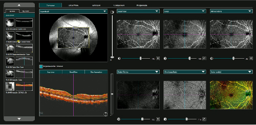
Optional VASCAN™ OCTA Module
*VASCAN™ OCT Angiography module is an optional software module of Mocean 4000.
Optical Coherence Tomography Angiography (OCTA) is a new non-invasive imaging technique that allows the detailed study of flow within the vascular structure of the eye without the need of dye injections.
· Ultraclear angiographic imaging powered by COMAG algorithm using phase and amplitude signals
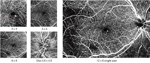
· Widefield OCTA imaging up to 12 mm x 8 mm
· SLO-based real-time retinal tracking effectively reduces motion artifacts and allows precise follow-up
· Advanced projection artifact removal algorithm ensures accurate visualization of the retinal vasculature
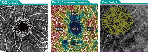
· Informative quantitative analysis including density, FAZ, impairment and flow analysis
· VASCAN provides a full view of the retina at 3x3, 6x6, 8x8 or 12x8, disc at 4.5x4.5 or 6x6
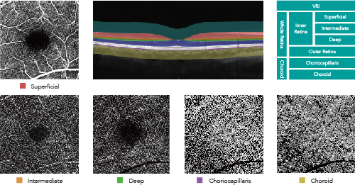
· VASCAN OCTA enables non-invasive vascular imaging of retinal vessels at 7 slabs and optic disc vessels at 4 slabs
For detailed information, please visit: OCTA Essential/Advanced
 中
中