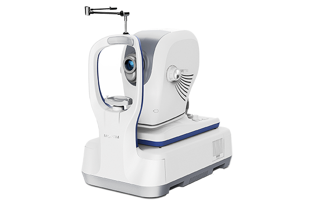


36,000 A-scans/s scanning speed

12mm wide scan for posterior segment

High quality real-time SLO

Eye tracker based on SLO retinal imaging

Deep Choroidal Imaging (DCI) mode reveals more details of the choroid

Comprehensive software analysis for retina, glaucoma and cornea
*Please contact us for availability in your country.


SLO-based retinal tracking
Eye tracking system uses Scanning Laser Ophthalmoscope (SLO) as a reference to minimize the involuntary eye movement during the image acquisition.

HD OCT and SLO imaging
During the scan, the system acquires up to 50 SLO images as well as 50 OCT images, then automatically generates high definition images using image averaging algorithm, which can effectively reduce speckle noise without compromising detail.
OCT IMAGING
Methodology
Spectral domain OCT
Optical source
Super luminescent diode (SLD), 840 nm
Scan speed
36,000 A-scans/s
Axial resolution (optical)
5 microns (optical), 2.7 microns (digital)
Transverse resolution
15 microns (optical), 3 microns (digital)
A-scan depth
2.1 mm
Diopter range
- 20 to + 20 diopters
Scan patterns
Macular: HD line scan (6 mm or 12 mm), 3D scan (6 mm x 6 mm), 6-line radial scan
Disc: 3D scan (6 mm x 6 mm)
Anterior: HD line scan (6 mm), 6-line radial scan
FUNDUS IMAGING
Methodology
Line scanning laser ophthalmoscopy (LSLO)
Minimum pupil diameter
3.0 mm
Field of view
47 degrees
SOFTWARE ANALYSIS
Macula
Retina thickness analysis; 3D view; En-face analysis; Progression analysis; EDI function
Glaucoma
RNFL analysis; Ganglion cell analysis; Cup-disk analysis; Progression analysis; OU comparative analysis
Anterior Segment
Manual measurement; Corneal thickness analysis
Others
DICOM conformance; Remote viewer software available
ELECTRICAL AND PHYSICAL
Weight
30.5 kg
Dimension
532 mm (L) x 360 mm (W) x 540 mm (H)
Source voltage
AC 100 - 240 V
Frequency
50 Hz - 60 Hz
Power input
90 VA