 中
中
- Company
-
Products
-
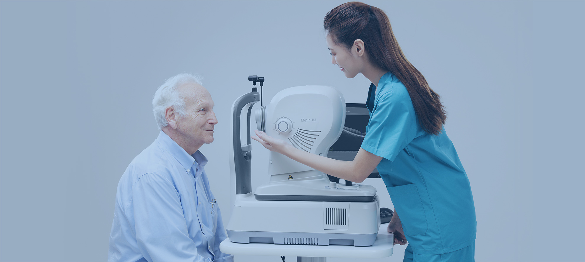
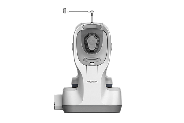
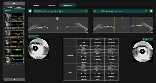
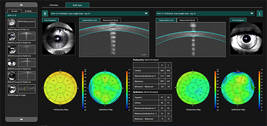
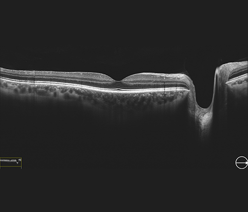
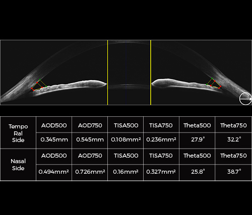
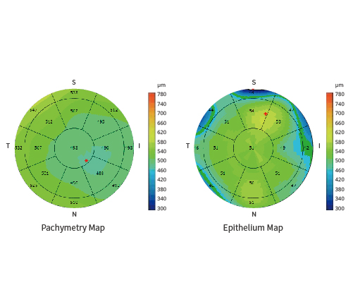
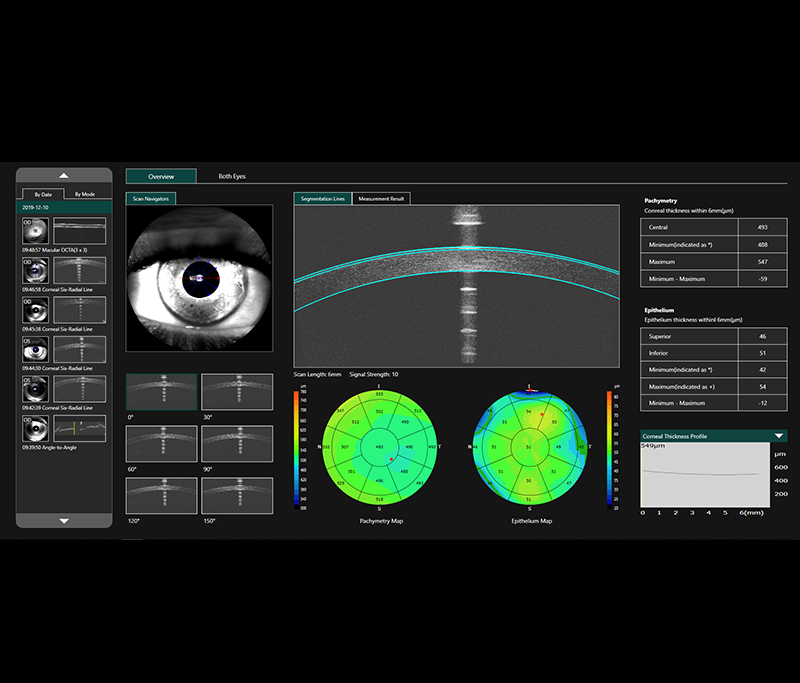
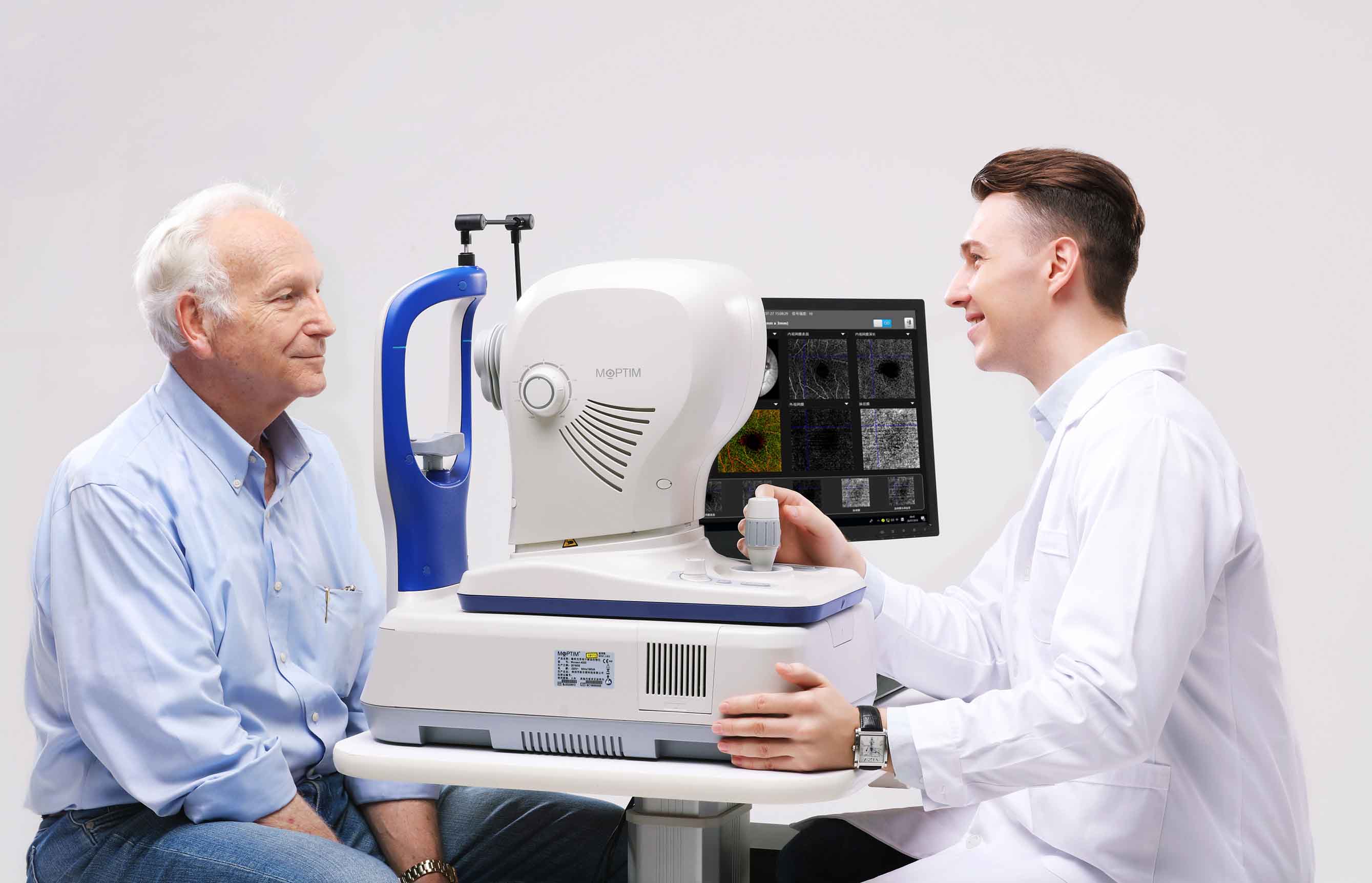
OCT/OCTA
Mocean 4000
Mocean 3000
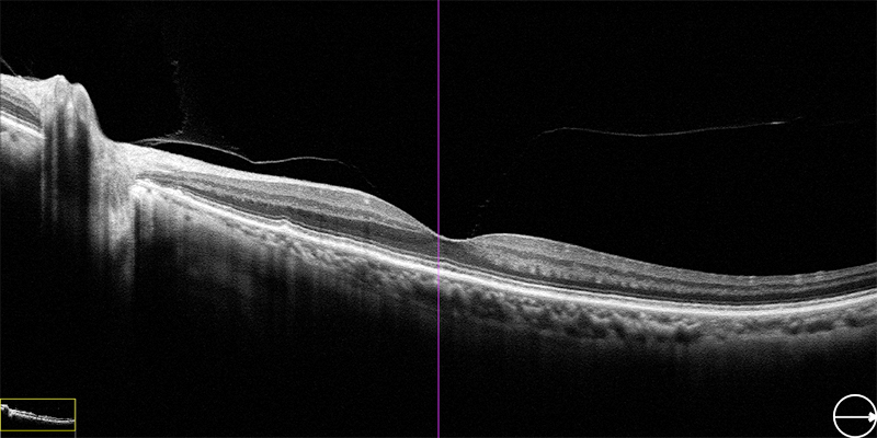
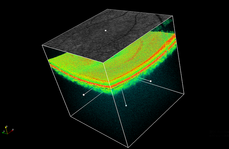
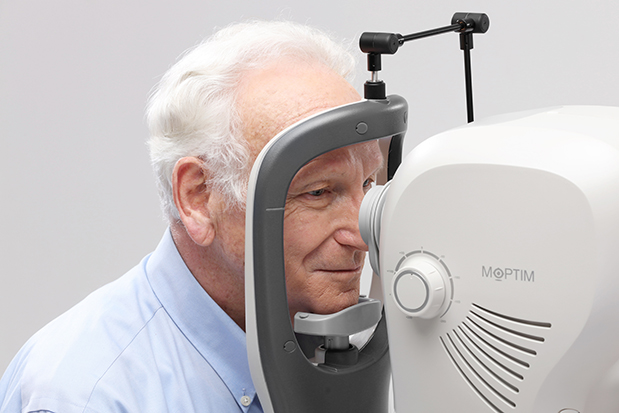
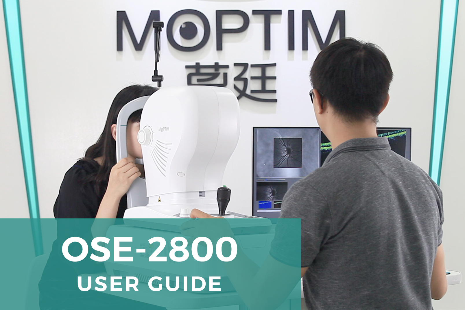
OSE-2800
-
Optical Biometer
Colombo IOL
Colombo IOL II
-
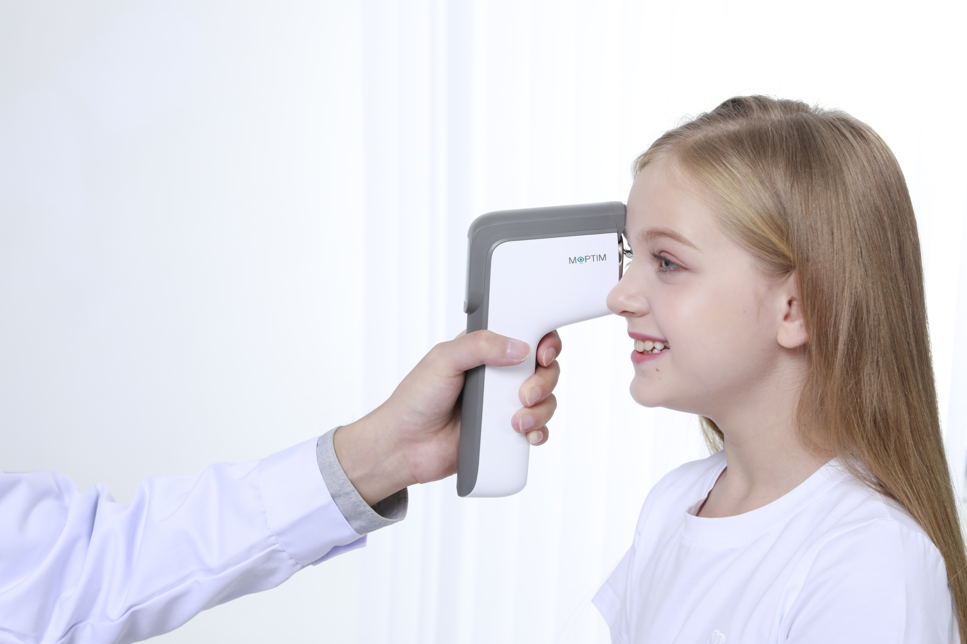
Dry Eye Analyzer
DEA
-
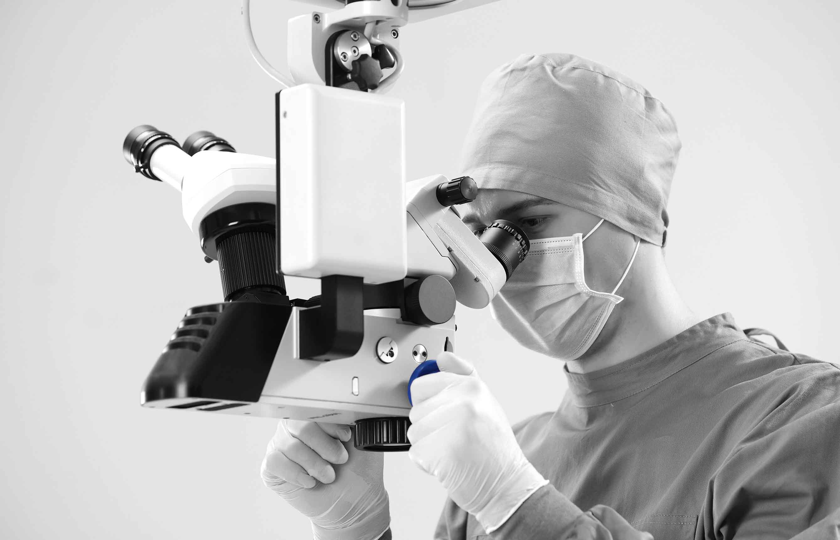
Operating Microscope
OPM 500
-
- References
- News & Events
- Distributors
- Supports


Shenzhen Certainn Technology Co., Ltd.
Member companies
Hangzhou Moptim Medical Equipment Co., Ltd.
Shenzhen certainn technology co., ltd. Zhengzhou branch
Shenzhen certainn technology co., ltd. Wuhan branch
Shenzhen certainn technology co., ltd. Chengdu branch
Shenzhen certainn technology co., ltd. Jinan branch
Shenzhen certainn technology co., ltd. Xian branch
Quick Links